


By Gisele Galoustian, FAU News Desk
Florida’s coral reefs are under siege. Since 2014, stony coral tissue loss disease (SCTLD) has spread rapidly across the Florida Reef Tract and Caribbean, killing vast numbers of reef-building corals and leaving behind dead skeletons where once-thriving reefs supported diverse marine life. Despite the severity of the crisis, little is known about how these diseases affect the microscopic structure of coral skeletons – the pores, densities and thicknesses that give reefs their strength and resilience.
Studying these tiny features has long been a challenge. Traditional methods are slow and often miss subtle structural changes.
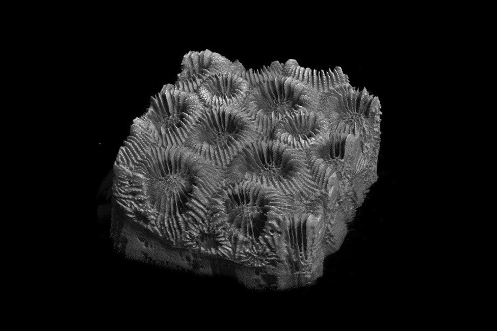
To address this challenge, Florida Atlantic University researchers turned to X-ray microcomputed tomography (micro-CT). The technique generates detailed 3D reconstructions down to microscopic pores, which reveal internal skeletal features, including porosity, thickness and structural orientation, in a non-destructive way. Housed in the FAU High School Owls Imaging Lab, the micro-CT was ideal for imaging corals, whose high mineral content provides strong X-ray contrast.
Researchers combined micro-CT imaging with deep learning-based image segmentation, using convolutional neural networks (CNNs), a form of artificial intelligence, to automatically distinguish coral skeletons from pore spaces. By analyzing images through patterns and features, this approach is faster and more accurate than traditional manual methods.
“Micro-CT gives us a window into the coral skeleton in a way that’s never been possible before,” said Alejandra Coronel-Zegarra, first author and a Ph.D. candidate in the Department of Chemistry and Biochemistry within FAU’s Charles E. Schmidt College of Science who won the 2025 Microscopy and Microanalysis Student Award for her research on SCTLD. “By combining it with deep learning, we can automatically detect subtle changes in the skeleton caused by disease – details that are nearly impossible to see manually.”
The team focused on two stony coral species: Montastraea cavernosa (M. cavernosa) and Porites astreoides (P. astreoides). By including both healthy and SCTLD-affected specimens, the researchers created a comprehensive dataset for testing the performance of several CNN models.
They investigated three U-Net-based deep learning models: U-Net, U-Net++, and Attention U-Net, known for capturing fine structural details. The models were trained to distinguish coral skeleton from pores and tested on four datasets, including healthy and SCTLD-affected M. cavernosa and healthy P. astreoides. Researchers tested how accurately each model detected subtle skeletal differences using standard metrics and statistical analysis.
Published in the Journal of Structural Biology, the results were striking. All three models performed exceptionally well, achieving more than 98% accuracy in distinguishing skeleton from pores.
“Without high-resolution, 3D insights, scientists cannot fully understand how disease, warming oceans and other stressors compromise reef survival,” said Vivian Merk, Ph.D., corresponding author and assistant professor in the Department of Chemistry and Biochemistry in FAU’s Charles E. Schmidt College of Science and the Department of Ocean and Mechanical Engineering in the College of Engineering and Computer Science. “Our analyses provide a clearer, quantitative picture of how environmental stressors reshape coral skeletons at the microscopic level. By uncovering these hidden changes in porosity, density and skeletal thickness, we can see exactly how diseases like Stony Coral Tissue Loss Disease alter the physical integrity of corals.”
Findings showed that Attention U-Net performed best, delivering high accuracy while working faster and across a range of coral species. It completed full image segmentation in just seven hours, compared to 15 hours for U-Net and 17 hours for U-Net++, making it especially useful for handling large, high-resolution micro-CT datasets.
Using these results, researchers created detailed 3D maps of coral skeletons. The analysis revealed clear differences between healthy corals and those affected by disease, showing how changes in pore structure may compromise skeletal integrity. Differences between species also emerged, highlighting how coral form and disease vulnerability are closely linked at the microscopic level.
“Beyond its immediate relevance to coral health, our research demonstrates the transformative potential of combining micro-CT with deep learning, and opens new possibilities for analyzing other biological materials, engineered composites and even geological samples,” said Merk. “This insight helps us identify reefs most at risk and develop more targeted protection and restoration strategies, strengthening the long-term resilience of Florida’s coral ecosystems.”
Study co-authors are Jamie Knaub, imaging lab assistant in FAU Lab Schools’ Owls Imaging Lab and a Ph.D. candidate in the FAU Department of Biology within the Charles E. Schmidt College of Science; and Abhijit Pandya, Ph.D., a professor in the Department of Electrical Engineering and Computer Science and Department of Biomedical Engineering within FAU’s College of Engineering and Computer Science.
This research was funded in part by the National Science Foundation awarded to Merk and seed funding from FAU’s College of Engineering and Computer Science and FAU’s I-SENSE Institute.
This piece was originally published at https://www.fau.edu/newsdesk/articles/stony-coral-imaging-study.php. Banner photo: Coral affected by stony coral tissue loss disease in the Carribean (G.Mannaerts, CC BY-SA 4.0, via Wikimedia Commons)
Sign up for The Invading Sea newsletter by visiting here. To support The Invading Sea, click here to make a donation. If you are interested in submitting an opinion piece to The Invading Sea, email Editor Nathan Crabbe.
